Osteoporosis Screening
Non irradiation sonogram testing
-
What is NO-radiation bone density screening?
-
Is a Dexa bone density scan dangerous? Why get a radiation Dexa Bone Density test when sonogram (non-radiation) bone density of the foot is available? Ask your doctor if the Achilles Bone Density can substitute for Dexa.
-
Can osteoporosis and osteopenia be reversed? Exercise of course counts, but reducing inflammation, a known contributor of bone loss, along with balancing hormone levels, improving nutrition and testing for malabsorption are essential for bone loss reversal.
Osteoporosis Screening
LISTEN TO DR. WALD’S RADIO SHOW ON EXERCISE HERE:
14. No Radiation Bone Density Test copyNo Radiation!!
Osteoporosis Screening – No Radiation!! Our method of bone density measurement uses the calcaneous – your heel bone. It is the most weight-bearing bone in the body. What’s best is that our technique uses sonogram (sound waves) and not x-ray radiation as conventional testing does, to accurately screen for loss of bone density (i.e., osteopenia and osteoporosis). Complementary medicine recognizes that degrading bone health may be secondary to absorption problems, chronic infections and disease, hormonal disorders and nutritional inadequacies. Osteoporosis screening is quick, painless and is part of a well-balanced nutritional and natural medicine approach. Please consult the information below and consider whether or not you should be tested.
Our bone densitometers is radiation free using high frequency sound waves (ultrasound) to evaluate bone status in the heel, the os calcis (the heal). The test is performed with the individual seated, with one foot placed on the foot positioner. The heel is surrounded by warm water encapsulated between inflated membranes. Water is the optimum medium for the transmission of ultrasound. A transducer on one side of the heel converts an electrical signal into a sound wave, which passes through the water and the individual’s heel. A transducer at a fixed distance on the opposite side of the heel receives the sound wave and converts it to an electrical signal that is analyzed. Our system measures the speed of sound (SOS) and the frequency-dependent attenuation of the sound waves (broadband ultrasound attenuation or BUA), and combines them to form a clinical measure called the Stiffness Index.
Strong bones allow for an active life!
Achilles Test for bone density (no radiation)
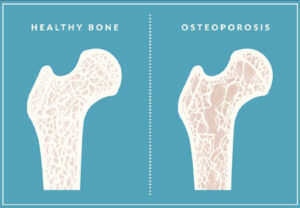
Osteoporosis is a condition where bones become weak to the point of breaking. This weakening may be due to aging, or caused by other factors that combine with age.
Important risk factors for osteoporosis include:
- Female
- Advanced Age
- An Existing Bone Fracture
- A Small Thin Frame
- Family History of Osteoporosis
- Removal of the Ovaries
- Early Menopause
- A Low Calcium Diet
- Lack of Exercise
- Eating disorders
- Certain Medications (Thyroid Medications, Steroids/Prednisones, Anticonvulsants, and Anticoagulants)
- Alcohol and Tobacco Use
According to WebMD –
 “Feb. 20, 2001 — New ultrasound devices that take measurements from the heel of the foot and do not use dangerous radiation are expected to put bone health checkups within the reach of many more patients and doctors. British researchers report in the February Journal of Bone and Mineral Research that “quantitative ultrasound,” or QUS, is as accurate as X-ray bone mineral density scans for identifying patients who are at risk for fractures within the next few years.”
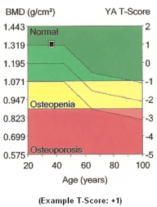
Who Should Have a Bone Density Test?
- NOF (The National Osteoporosis Foundation) recommends that you have a bone density test if:
- you are a woman age 65 or older
- you are a man age 70 or older
- you break a bone after age 50
- you are a woman of menopausal age with risk factors
- you are a postmenopausal woman under age 65 with risk factors
- you are a man age 50-69 with risk factors
- A bone density test may also be necessary if you have any of the following:
- an X-ray of your spine showing a break or bone loss in your spine
- back pain with a possible break in your spine
- height loss of ½ inch or more within one year
- total height loss of 1½ inches from your original height
Scientific References
References:
Heel Ultrasound as a Predictor of Appendicular Bone Mineral Density – http://analytics.ncsu.edu/sesug/2001/P-716.pdf
1. International Osteoporosis Foundation. 2. Zagzebski JA, Rossman PJ, Mesina C, Mazess RB, and Madsen EL. (1991) Ultrasound transmission measurements through the os calcis. Calcif Tissue Int 49:107–111. 3. Rossman P, Zagzebski J, Mesina C, Sorenson J, Mazess R. (1989) Comparison of ultrasonic velocity and attenuation in the os calcis to photon absorptiometry measurements in the radius, femur, and lumbar sine. Clin Phys Physiol Meas 10:353–360. 4. Langton CM, Palmer SB, and Porter RW. (1984) The measurement of broadband ultrasonic attenuation in cancellous bone. Eng Med 13:89–91. 5. Hans D, Schott AM, and Meunier PJ. (1993) Ultrasonic assessment of bone: a review. Eur J Med 2(3):157–163. 6. Hans D, Fuerst T, Duboeuf F. (1997) Quantitative ultrasound bone measurement. Eur Radiol 7 (Suppl 2):S43–S50. 7. Bouxsein ML, Radloff SE. (1997) Quantitative ultrasound of the calcaneus reflects the mechanical properties of calcaneal trabecular bone. J Bone Miner Res 12:839– 846. 8. Han S, Medige J, Davis J, Fishkin Z, Mihalko W, Ziv I. (1997) Ultrasound velocity and broadband attenuation as predictors of load-bearing capacities of human calcanei. Calcif Tissue Int 60:21–25. 9. Duquette J, Lin J, Hoffman A, Houde J, Ahmadi S, Baran D. (1997) Correlations among bone mineral density, broadband ultrasound attenuation, mechanical indentation testing, and bone orientation in bovine femoral neck samples. Calcif Tissue Int 60:181–186. 10. Njeh CF, Hodgskinson R, Currey JD, Langton CM. (1996) Orthogonal relationships between ultrasonic velocity and material properties of bovine cancellous bone. Med Eng Phys 18:373–381. 11. Langton CM, Njeh CF, Hodgskinson R, Currey JD. (1996) Prediction of mechanical properties of the human calcaneus by broadband ultrasonic attenuation. Bone 18:495– 503. 12. Hodgskinson R, Njeh CF, Whitehead MA, Langton CM. (1996) The nonlinear relationship between BUA and porosity in cancellous bone. Phys Med Biol 41:2411– 2420. 13. Laugier P, Droin P, Laval-Jeantet AM, Berger G. (1997) In vitro assessment of the relationship between acoustic properties and bone mass density of the calcaneus by comparison of ultrasound parametric imaging and quantitative computed tomography. Bone 20:157–165. 14. Tavakoli MB and Evans JA. (1991) Dependence of the velocity and attenuation of ultrasound in bone on the mineral content. Phys Med Biol 36:1529–1537. A-6 Theory of Ultrasound Achilles InSight/Express Operator’s Manual 15. Evans JA and Tavakoli MB. (1990) Ultrasonic attenuation and velocity in bone. Phys Med Biol 35:1387– 1396. 16. Hans D, Arlot ME, Schott AM, Roux JP, Kotzki PO, Meunier PJ. (1995) Do ultrasound measurements on the os calcis reflect more the bone microarchitecture than the bone mass?: a two-dimensional histomorphometric study. Bone 16:295–300. 17. Gluer CC, Vahlensieck M, Faulkner KG, Engelke K, Black D, and Genant HK. (1992) Sitematched calcaneal measurements of broad-band ultrasound attenuation and single x-ray absorptiometry: do they measure different skeletal properties? J Bone Miner Res 7:1071–1079. 18. Nicholson PHF, Haddaway MJ, Davie MWJ. (1994) The dependence of ultrasonic properties on orientation in human vertebral bone. Phys Med Biol 39:1013–1024. 19. Clarke AJ, Evans JA, Truscott JG, Milner R, Smith MA. (1994) A phantom for quantitative ultrasound of trabecular bone. Phys Med Biol 1677–1687. 20. Krieg M-A, Barkmann R, Gonnelli S, et al. Quantitative Ultrasound in the Management of Osteoporosis: The 2007 ISCD Official Positions. J Clin Densitom: Assessment of Skeletal Health. 2008; 11 (1): 163-187. 21. Simon D, Boring III JR. Chap. 6: Sensitivity, Specificity, and Predictive Value. In: HK Walker, WD Hall, JW Hurst, ed. Clinical Methods: The History, Physical, and Laboratory Examinations, 3rd edition. Boston: Butterworths; 1990. 22. Gonnelli S, Cepollaro C, Pondrelli C, Martini S, Rossi S, Gennari C. Ultrasound parameters in osteoporotic patients treated with salmon calcitonin: A longitudinal study. Osteoporos Int. 1996; 6 (4): 303-307. 23. Giorgino R, Paparella AP, Lorusso D, Mancuso S. Effects of oral alendronate treatment and discontinuance on ultrasound measurements of the heel in postmenopausal osteoporosis. North American Menopausal Society, September 1996. 24. Gonnelli S, Cepollaro C, Montagnani A, et al. Heel ultrasonography in monitoring alendronate therapy: a four-year longitudinal study. Osteoporos Int. 2002; 13 (5): 415-421.
 Previous Post
 Next Post